★ Win a pair of Brandecosse Capriolo boots worth £219! Click here to enter ★

What you need to know about hip dysplasia and gundogs
 Labrador
Usd 5 dec 18 vintage times
Labrador
Usd 5 dec 18 vintage times
A good question and well done for admitting that you don’t know about it or understand what hip dysplasia in dogs is.
Explaining hip dysplasia in dogs
The hip joints are at the top of the hind leg in dogs and are ‘ball and socket’ joints, so that flexion (lifting the knee up and forward), extension (moving the straight leg back the same way) and rotation (think squatting to pee) are all possible.
The joint is formed by the round ‘head’ of the femur (or thigh bone) inserting into the acetabulum of the pelvis, which is the ‘socket’. Some stability is added by the presence of the ‘round’ ligament, which attaches the femoral head to the acetabulum, and the joint capsule that surrounds the joint.
What we would want in an individual is a nice, round ball fitting snugly into a nice, round socket. But, of course, sometimes things can go wrong.
Why hip dysplasia in dogs occurs
Despite what some breeders will tell you, hip dysplasia is considered an inherited trait. However, it is also influenced by environmental factors.
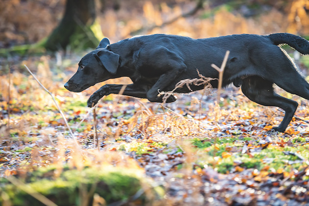
- Size and breed: Particular breeds are associated with the disease, so that, for example, it is far more common in Labradors than in spaniels.
- Growth rate: Exaggerated growth rates contribute towards overloading of the hip. Puppies should be fed according to growth charts and adult weight should not be achieved until they are adults. We really do not want to see fat puppies.
- Type of feeding: Minerals and vitamins should be balanced with calorie intake, so that a puppy of normal body weight will be correctly mineralised. This is difficult or impossible to achieve with most home-made diets. Diets high in meat will inevitably not possess the correct calcium:phosphorus ratio.
- Type and duration of exercise: Little and often is required. Puppies should not be ‘stretched’ by running them with stronger, faster adult dogs. Avoid jumping and concentrate on lead work instead.
Severe hip dysplasia with significant remodelling of the joint. This dog was sound with only restricted movement evident.
The problem
If in a young, growing animal, the soft tissue surrounding the joint fails to support it properly, then laxity of the joint will occur. Depending on the degree of movement, a destructive chain of events follows:
• Inflammation in the joint capsule causes pain
• Joint fluid increases, further destabilising the joint
• The bones move together abnormally causing erosive damage
• The round ligament is stretched allowing even more abnormal movement
• Lameness results in poor muscle development
• Tiny fragments of bone float in the joint (joint mice)
• Remodelling of the femoral head and neck occur
• The femoral head flattens and the neck thickens
• In less severe cases, stability returns but movement is restricted
• Further deterioration after maturity
Clinical signs
Clinical signs of hip dysplasia in dogs are usually obvious but stalwart dogs can appear normal until their gait is examined closely. Affected dogs may be reluctant or slow to rise, climb stairs and/or jump. Intolerance of exercise is common. There is pain on extension of the hip, especially when it is also rotated outwards. Well-muscled forequarters can cast some suspicion, as weight is thrown forward to protect the hip. When both hips are affected, you may see a ‘bunny hop’ gait and a ‘kyphotic’ posture (forward rounding of the lower back). Diagnosis can be confirmed by X-ray, which must be done under general anaesthesia or heavy sedation, so that luxation (dislocation) of the hips can be assessed and specific tests (Barden and Ortolani signs) be undertaken. It must be noted, however, that there is no correlation between the severity of the radiological changes and the degree of lameness. Some working dogs seem pretty resistant to the effects of hip dysplasia but care should be taken that these individuals are not bred from. And no matter what anyone says, it is just not possible to assess the quality of the hip joint without X-rays.
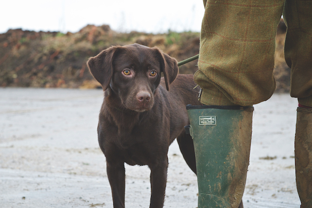
Correct vitamin and mineral uptake is vital in developing dogs
Prevention
Sometimes you need a bit of luck, but it helps to source pups from tested parents that have good ‘hip scores’. The BVA Hip Dysplasia Scheme calculates a score for each hip. Most dogs require anaesthesia to allow a perfectly straight ventro-dorsal X-ray of the hip joints to be taken. The best score per side is 0 and the worst 53. Thus perfect hips score 0-0. Only dogs with better than Breed Mean Score hips should be bred from. In Labradors, this would be significantly better than a total score of 18. The results for individual dogs can be viewed on the Kennel Club’s website.
The outcome
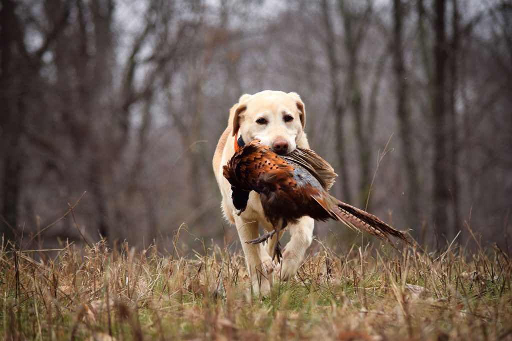
Many affected young dogs will, with conservative management, weight loss and anti-inflammatory treatment, return to being sound but they will rarely be able to work successfully. In older dogs, progressive arthritis generally occurs, requiring medical input.
Related Articles
The vital role of gundogs in driven game shooting
For many, what makes the shoot day all the better is the company of our dogs, whether on the peg or beating line, says Julia Newman.
By Shooting Times
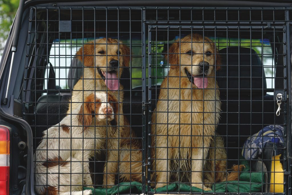
Safe gundog travel: why you must restrain your dog in the car
Discover why properly restraining your gundog in the car is essential for safety, legality, and insurance. Learn about crash-tested crates, harnesses and Highway Code rules for travelling with dogs.
By Shooting Times
Get the latest news delivered direct to your door
Subscribe to Shooting Times & Country
Discover the ultimate companion for fieldsports enthusiasts with Shooting Times & Country Magazine, the UK’s leading weekly publication that has been at the forefront of shooting culture since 1882. Subscribers gain access to expert tips, comprehensive gear reviews, seasonal advice and a vibrant community of like-minded shooters.
Save on shop price when you subscribe with weekly issues featuring in-depth articles on gundog training, exclusive member offers and access to the digital back issue library. A Shooting Times & Country subscription is more than a magazine, don’t just read about the countryside; immerse yourself in its most authoritative and engaging publication.